 |
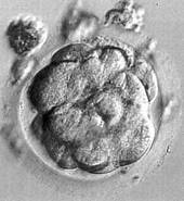
After about two and a half days the embryo, by now at about the 8-cell stage enters the uterus. By then a remarkable and significant change has occurred. The spherical blastomeres flatten against each other producing a larger area of cell-to-cell contact, and communication between the blastomeres begins. This process is called compaction, and the embryo is now known as a morula. The significance of this stage is that for the first time some cells of the embryo are different from others; this marks the beginning of the process of differentiation. The cells of the embryo are no longer totipotent.
|
 | |
Blastocyst |
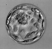
A fluid filled cavity appears within the cells of the morula, and as the cells continue to divide, the cavity enlarges, eventually producing a hollow sphere consisting of two distinct types of cell. This structure is called a blastocyst. It consists of a single layer of flattened cells, the trophectoderm, which is destined to become the placenta and amniotic membranes and a group of smaller cells, called the inner cell mass, which is destined to become the foetus itself.
 Task:
Task:
Print out and label the picture of the blastocyst: zona pellucida, outer layer of cells, inner cell mass, fluid filled cavity.
 | |
A hatching blastocyst |
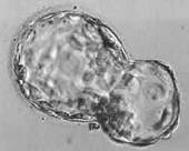
Up to this point, the zona pellucida has served to protect the developing embryo, but now it will get in the way of the embryo embedding itself in the wall of the uterus, where it will continue to grow and develop until birth. By about 5 days after fertilisation, enzymes break down the zona pellucida, and the blastocyst is able to "hatch" from it by squeezing through a small hole.
Implantation is when for the first time the embryo comes into intimate association with the circulation of the mother, to ensure its survival. Now free of its entire original protective layers, the blastocyst sticks to the lining of uterus, which has become specially thickened to receive it. The blastocyst embeds itself in the lining and the outer layer cells continue to divide and differentiate into the placenta and membranes, whilst the inner cell mass undergoes an extraordinary pattern of genetically programmed growth and development to become a foetus.
![]() Review the stages of development from fertilisation to implantation and visit again the question of when life itself might begin.
Review the stages of development from fertilisation to implantation and visit again the question of when life itself might begin.


What's your opinion?
Average rating


Not yet rated